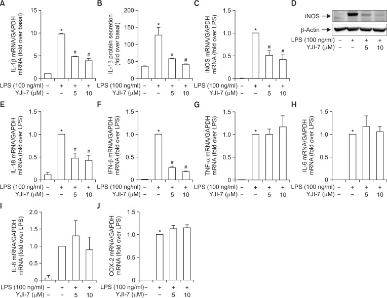
Fig. 5.
Effects of YJI-7 on the expression of inflammatory mediators in RAW 264.7 macrophages. (A, B) RAW 264.7 macrophages were pretreated with indicated concentration of YJI-7 for 2 h prior to stimulation with LPS. (A) IL-1β mRNA expression was measured by qRT-PCR and normalized to that of GAPDH as described in Materials and Methods. (B) Secreted IL-1β was measured by ELISA. Values are presented as fold change compared to LPS-stimulated cells and are expressed as the mean ± SEM (n=3). *p<0.05 compared with control group, #p<0.05 compared with cells treated with LPS only. (C, D) Cells were pretreated with YJI-7 for 2 h followed by the incubation with LPS. (C) iNOS mRNA expression level was determined by qRT-PCR and normalized to that of GAPDH. Values are represented as fold change compared to LPS-stimulated cells and are expressed as mean ± SEM (n=3). *p<0.05 compared with control group, #p<0.05 compared with cells treated with LPS only. (D) Protein expression of iNOS was measured by Western blot analysis. (E-J) Cells were treated with YJI-7 for 2 h followed by the stimulation with LPS. Expression levels of IL-18 (E), IFN-β (F), TNF-α (G), IL-6 (H), IL-8 (I) and COX-2 (J) mRNA were determined by qRT-PCR. Values are presented as fold change compared to LPS-stimulated cells and are expressed as mean ± SEM (n=3). *p<0.05 compared with control group, #p<0.05 compared with cells treated with LPS only.

