Fig. 11.

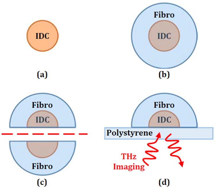
Steps in phantom tumor model formation with (a) IDC phantom core, (b) phantom fibro surrounding IDC, (c) tumor model bisection, and (d) THz reflection imaging setup where the emitter and detector are below the sample.

Steps in phantom tumor model formation with (a) IDC phantom core, (b) phantom fibro surrounding IDC, (c) tumor model bisection, and (d) THz reflection imaging setup where the emitter and detector are below the sample.