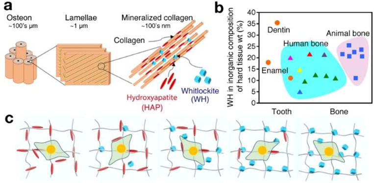
Fig. 1.

The two major inorganic components of bone: hydroxyapatite (HAP: Ca10(PO4)6(OH)2) and whitlockite (WH: Ca18Mg2(HPO4)2(PO4)12) nanocrystallites. a) Schematic of bone structure ranging from the nanometer to micrometer scales, showing that bone tissue consists of cylindrical osteon units at the microscale, which are composed of collagen nanofibers with HAP and WH bone mineral particles at the nanoscale. b) Amount (weight percent) of WH in the inorganic part of hard tissues, calculated based on the magnesium amount. Data were obtained from previous publications, with different colors representing different references (Orange circle: Driessens et al. [16], Magenta triangle: Gabriels et al. [10], red triangle: Carlstrom et al. [16], purple triangle: Breibart et al. [11], blue triangle: Wooddard et al. [12], yellow triangle: Duckworth et al. [13], green triangle: Schraer et al. [14], blue rectangle: Long et al. [15] c) Schematic of experiments to identify optimal conditions in the stem cell niche for osteogenic differentiation of human mesenchymal stem cells (MSCs) with regard to the different ratios of HAP and WH.
