Fig. 2.

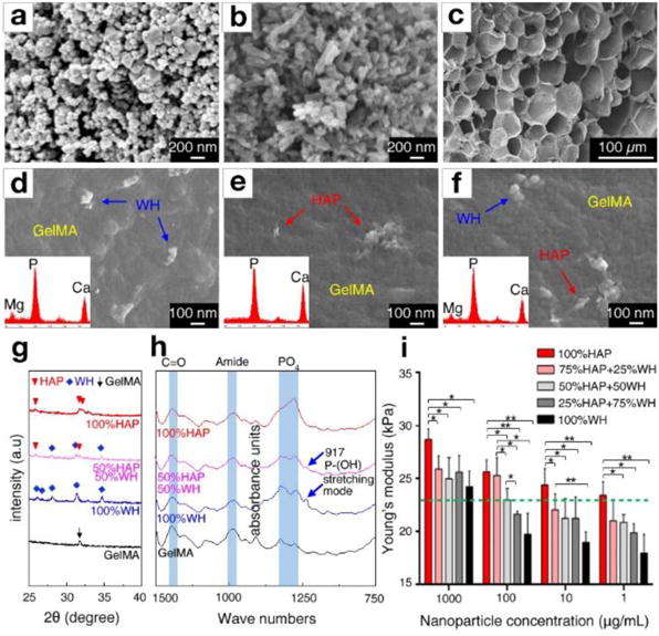
Engineered bone-mimetic nanocomposite hydrogel scaffolds. FESEM observation of a) synthesized WH nanoparticles with rhombohedral shape, b) HAP nanoparticles with needle shape, and c) microporous structured GelMA hydrogel scaffold after freeze drying. d-f) The nanoscale features of composite hydrogel scaffolds, showing that rhombohedral shaped WH nanoparticles and needle-shaped HAP nanoparticles are homogeneously dispersed in the GelMA hydrogel. Insets in each panels show the EDS area scan analysis, where WH incorporated GelMA hydrogel had a Mg peak, and HAP incorporated GelMA exhibited a higher ratio of the Ca and P peak intensity. g) XRD analysis of HAP-incorporated GelMA hydrogel and WH-incorporated GelMA hydrogel, confirming that their crystal phase is maintained after fabrication. h) FT-IR analysis result of HAP-incorporated GelMA hydrogel and WH-incorporated GelMA hydrogel, confirming that the chemical groups of HAP, WH and GelMA hydrogel remain intact after fabrication. i) The Young’s modulus of the composite hydrogel scaffolds depending on different concentrations and ratios of HAP and WH in GelMA hydrogel were calculated from compression tests. The green dotted line indicates the stiffness of pure GelMA hydrogel (~23 kPa). (*p<0.05, **p<0.01)
