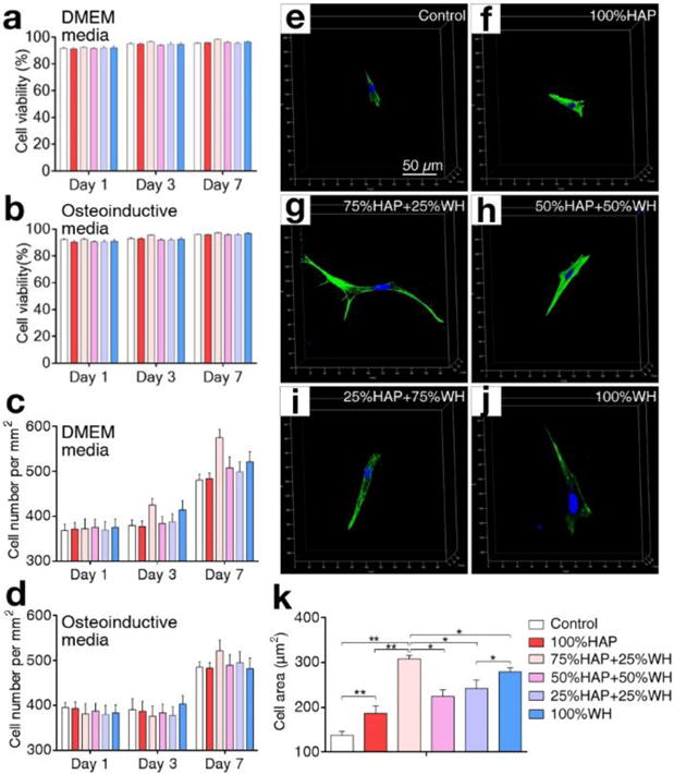
Fig. 3.

Cellular viability, proliferation, and spreading in composite hydrogel scaffolds with different ratios of HAP and WH. a–d) Viability (a–b) and proliferation (c–d) of cells grown in 3D composite hydrogel scaffolds with different ratios of HAP and WH, in two media conditions (DMEM and osteoinductive media) were compared. e–k) Spreading of cells in 3D composite hydrogel scaffolds depending on different ratios of HAP and WH at day 7 was observed under confocal microscopy and quantified based on their spreading area. Cellular nuclei and actin were stained with DAPI (blue) and phalloidin (green). (*p<0.05, **p<0.01)
