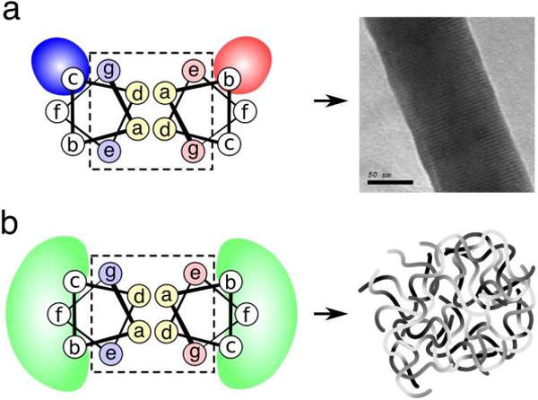
Fig. 3.

(a) In previous SAF designs, the solvent exposed residues b and c contained specific charged residues, which led to helix alignment and thick fiber formation. (b) In hSAF designs, the b, f, and c positions were changed to combinations of glutamine and alanine with the goal of achieving weaker interactions, leading to small, flexible bundles of fibers
