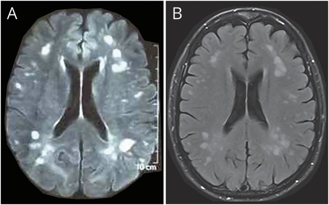
Figure. Fluid-attenuated inversion recovery (FLAIR) MRI.

(A) FLAIR MRI taken 36 hours after the onset of symptoms and before methylprednisolone therapy shows innumerous, asymmetric, patchy, and poorly marginated areas of increased signal intensity. (B) FLAIR MRI taken 2.5 months after the onset of symptoms shows residual lesions, but no new lesions.
