Figure 4.
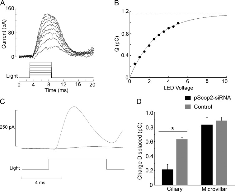
pScop2 is photoisomerized by light and underlies the ERC. (A) ERCs evoked by flashes of increasing intensity (470 nm, 6 ms in duration, 1.3 × 10¹⁷ to 9.1 × 10¹⁷ photons ⋅ s−1 ⋅ ⋅cm−2) in a ciliary photoreceptor voltage-clamped at −40 mV. Between trials, a 5-s period of red illumination (620 nm) was interposed 1 min before the subsequent test flash, to reset the photopigment to the R state. (B) Saturation of the charge displaced by illumination. The amount of charge displaced at each stimulus intensity was obtained by integrating the corresponding current traces of A. An exponential function was least-squares fitted to the data points to extract the estimated asymptote; this represents the maximum photoisomerization charge, which in turn reflects the total amount of photopigment in the cell (r2 = 0.992). (C) Comparison of the ERC obtained in a control ciliary photoreceptor and one previously treated with pScop2-siRNA; the treatment greatly attenuated the ERC. Test flash, 7.9 × 10¹⁷ photons ⋅ s−1 ⋅ cm−2, 6-ms duration. (D) Mean total photoisomerization charge under control and RNAi conditions, for the two photoreceptor types in the double-retina of Pecten. Before dissociation and electrophysiological testing, the retinae were subjected to electroporation in the presence of either pScop2-siRNA or fluorescent dextran as a control and cultured for 24–48 h. Only in ciliary photoreceptors did the RNAi treatment depress the amount of photoisomerization charge (*, P < 0.01, t test). Error bars indicate SEM.

