Figure 3.
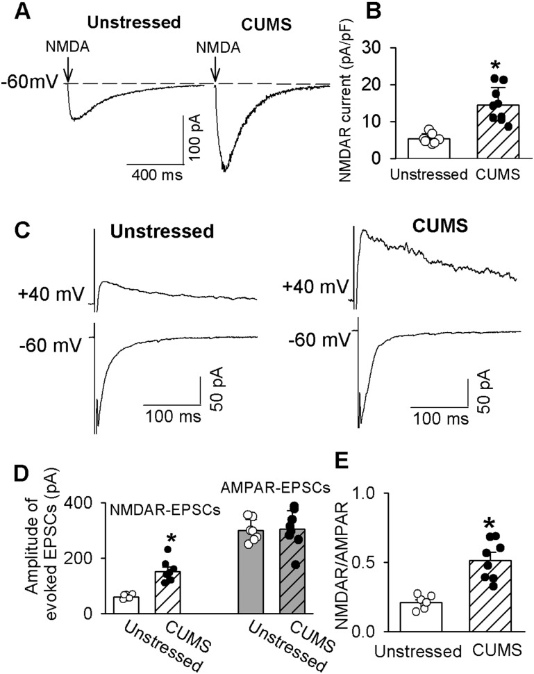
CUMS enhances synaptic NMDAR activity in PVN-CRH neurons. (A) Original current traces and (B) summary data show currents elicited by puff 100 µM NMDA in eGFP-tagged PVN-CRH neurons from CUMS (n = 9 neurons) and unstressed rats (n = 7 neurons). (C) Representative traces and (D) summary data of evoked AMPAR-EPSCs (holding potential of −60 mV) and NMDAR-EPSCs (holding potential of 40 mV) in eGFP-labeled neurons from CUMS rats (n = 8 neurons) and unstressed rats (n = 7 neurons). (E) Group data show the ratios of NMDAR-EPSCs to AMPAR-EPSCs in neurons from unstressed and CUMS rats in D. *P < 0.05 compared with unstressed rats.

