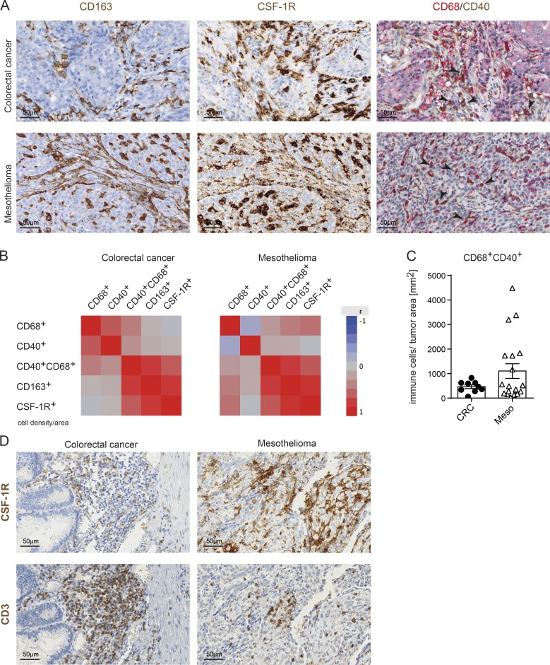
Figure 5.
CD40 is expressed on TAMs of cancer patients. (A) Exemplary IHC images for CD163 and CSF-1R staining and duplex staining for CD68/CD40 staining of one exemplary CRC and one exemplary mesothelioma patient. Black arrowheads indicate examples of CD68+CD40+ double-positive macrophages. (B) Overall correlation shown as heat maps of 9 CRC and 19 mesothelioma patients using data obtained by automated digital analyses. (C) CD68+CD40+ double-positive cells in CRC and mesothelioma patients quantified as cell counts per squared millimeters of tumor tissue by digital analyses. (D) CSF-1R+ macrophages and CD3+ T cells colocalize in CRC and mesothelioma as assessed from consecutive sections.

