Figure 6.
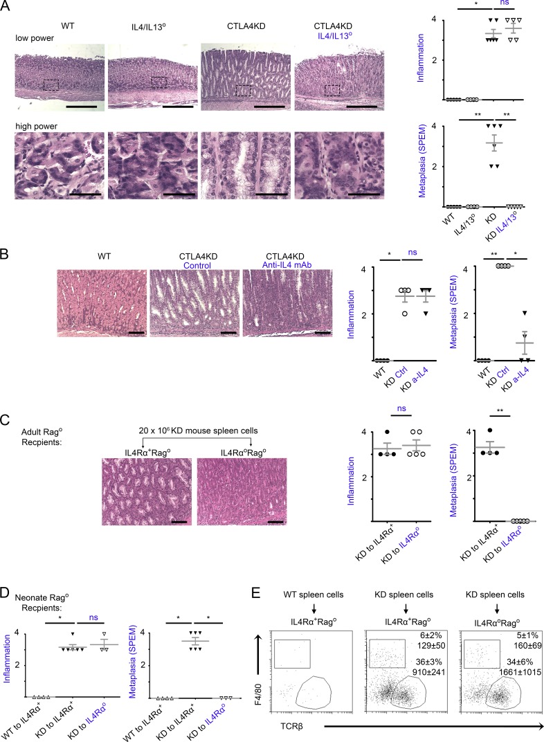
IL-4 and IL-13 were required for initiation of inflammatory tumorigenesis by CTLA4 insufficiency. (A) CTLA4KD IL4/IL13° mice (knockout of IL-4 and adjacent IL-13 genes) were analyzed at 8–10 wk of age in comparison to three types of littermate controls: wild-type, IL4/IL13°, and CTLA4KD(IL4/IL13+). Data represent four to six mice per group pooled from two experiments (mean ± SEM; Kruskal–Wallis test). Bars: (low power) 500 µm; (high power) 50 µm. (B) CTLA4KD mice were treated with anti–IL-4 mAb for 4 wk and analyzed at 5–6 wk of age. Data represent four mice per group pooled from two experiments (mean ± SEM; Kruskal–Wallis test). Bars, 100 µm. (C and D) IL4RαoRago mice or IL4Rα+Rago controls at 6 wk (C) or 2–3 d of age (D) were reconstituted with 20 × 106 splenocytes from CTLAKD or normal mice and analyzed 5–6 wk later. Representative gastric histopathology images (bars, 100 µm) are followed with a summary of pathology scores. Data represent three to six mice per group pooled from two experiments. Each data point represents one animal (mean ± SEM; Mann–Whitney test in C and Kruskal–Wallis test in D). (E) Flow cytometry analyses of IL4RαoRago and IL4Rα+Rago mice reconstituted with spleen cells (groups shown in D) for immune cell profile in the stomach mucosa. The plots are gated on live cells (identified by viability dye), showing percentage of αβ T cells and macrophages of the CD45+ population, and the total number of the gated population recovered from approximately one fourth of the stomach mucosa used for flow cytometry. *, P < 0.05; **, P < 0.01; ns, not significant.

