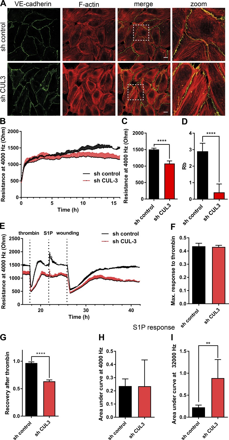
Figure 1.
Knockdown of Cullin-3 impairs cell signaling involved in endothelial barrier maintenance. (A) HUVECs were transduced with the control shRNA or Cullin-3–targeting shRNA and stained for VE–cadherin and F-actin at 72 h after infection. Dashed boxes correspond to zoomed images. Bars, 15 µm. (B) ECIS measurement of HUVECs prepared as in A. 105 cells were seeded per well in an eight-well ECIS slide at 72 h after infection, and electrical resistance was measured at 4,000 Hz (n = 5). (C) Resistance values at 4,000 Hz were compared by analysis of 10 measurement points 16–17 h after seeding for the cells prepared as in A (n = 5). (D) Quantification of the Rb parameter was analyzed as in C. (E) HUVECs were prepared as in A, and the resistance of the endothelial monolayer was measured at 4,000 Hz using ECIS. Cells were stimulated with 1 U/ml thrombin and, after recovery, with 500 nM S1P, and electrical wounding was performed at 100 kHZ, 6,500 µA for 120 s. (F) Maximum response to thrombin response was calculated using GraphPad Prism (n = 5). (G) Recovery upon thrombin stimulation was measured at 1.5 h upon thrombin addition and normalized to the resistance values before thrombin addition (n = 5). (H and I) Analysis of S1P response in cells prepared as in A (n = 5). Areas under the curve of S1P response were calculated using GraphPad Prism. Error bars represent SD. **, P = 0.01–0.001; ****, P < 0.0001.

