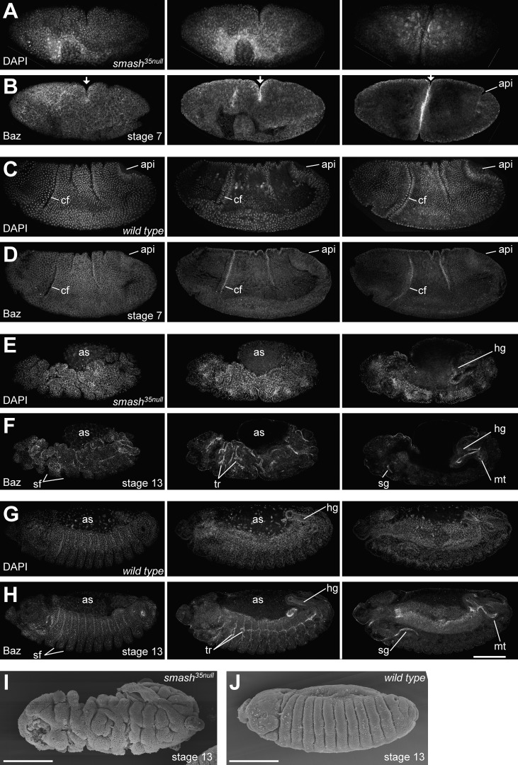
Figure 6.
Embryos lacking maternal and zygotic Smash show severe defects in morphogenesis. (A–H) smash35null embryos at stage 7 (A and B) and stage 13 (E and F) stained for DNA (DAPI) and Baz were compared with WT embryos at the corresponding stages (C, D, G, and H). Three different optical sections of the same embryo are shown for each stage. The left column shows the most superficial optical sections, whereas the middle and right columns show deeper optical sections to visualize internal organs. The mutant embryo in A and B lacks the cephalic furrow (cf) and instead has formed a deep ectopic furrow in the middle (arrows). It also fails to form a proper amnioproctodeal invagination (api). The mutant embryo in E and F has a very irregular shape, with deep clefts in its surface. Segmental furrows (sf) are irregular in shape, position, and depth. Morphogenesis of tubular organs such as hindgut (hg), Malpighian tubules (mt), salivary glands (sg), and tracheae (tr) is highly abnormal. The yolk covered by the amnioserosa (as) bulges out of the dorsal side of the embryo. (I and J) Scanning EM of smash35null (I) and WT (J) embryos at stage 13. Note the extremely irregular surface structure of the smash35null embryo in I. Bars, 100 µm. Anterior is to the left and dorsal is up. The full z-stacks of A–H are shown in Videos 1–4.

