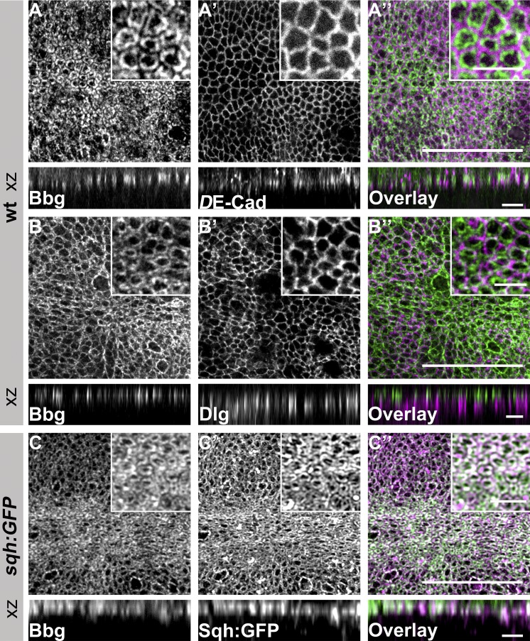
Figure 3.
Bbg localizes in the apical cytocortex of L3 wing disc epithelial cells. (A–B′′) WT L3 wing discs stained with anti-Bbg (A and B), anti–DE-Cadherin (DE-Cad; A′), anti-Dlg (B′), and the respective overlays (A′′ and B′′). (C–C′′) sqh-GFP L3 wing disc stained with anti-Bbg (C), Sqh-GFP (endogenous signal, C′) and the respective overlay (C′′). The projection in B was taken from a more lateral view compared with that of A and C. Insets, top right: Respective pouch areas. xz projection shows the central area of the same L3 wing discs. Bars: (A′′, B′′, and C′′) 25 µm; (xz projections and small boxes) 5 µm.

