Figure 2.
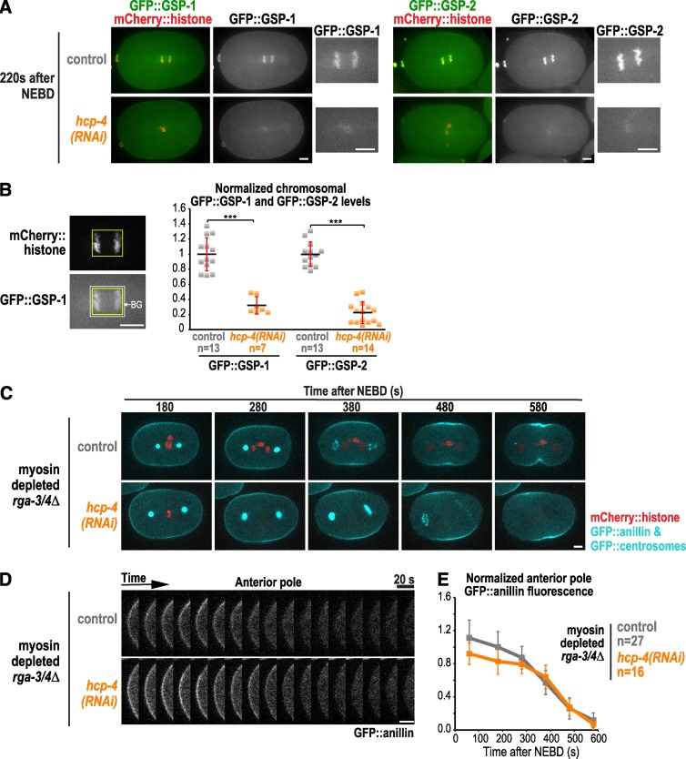
Kinetochore-localized PP1 is not required for polar clearing. (A) Representative images of control (n = 13) and hcp-4(RNAi) embryos expressing mCherry::histone and in situ fusions of GFP with GSP-1 (left; n = 7) or GSP-2 (right; n = 14). (B) Mean chromosomal GFP::GSP-1 and GFP::GSP-2 fluorescence was measured 200 s after NEBD. n = number of embryos; error bars are the SD; p-values are two-tailed Student’s t test (***, P < 0.001). (C) Time-lapse series of myosin-depleted rga-3/4Δ embryos expressing GFP::anillin, a GFP::centrosome marker (GFP::SPD-5) and mCherry::histone. Representative control (top; n = 14) and hcp-4(RNAi) embryos (bottom; n = 9) are shown. (D) Kymographs of the anterior pole in the embryos in C beginning 180 s after NEBD. (E) Normalized cortical GFP::anillin fluorescence is plotted for the anterior pole. The control data are reproduced from Fig. 1 G for comparison. n = number of linescans; error bars are SEM. Bars, 5 µm.

