Figure 5.
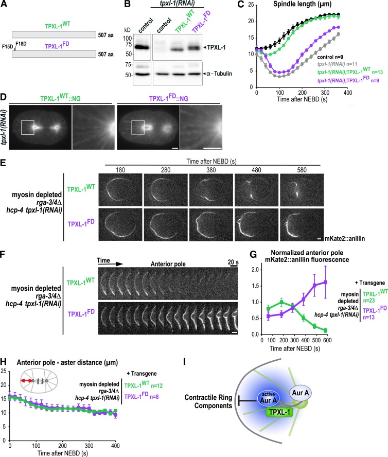
Polar clearing requires the ability of TPXL-1 to activate Aurora A. (A) Schematics of the protein products of the WT and Aurora A–binding defective (FD) tpxl-1 transgenes. (B) Immunoblots of control (N2) worms and worms expressing TPXL-1WT or TPXL-1FD after depletion of endogenous TPXL-1 by RNAi were probed for TPXL-1 and α-tubulin as a loading control. (C) Spindle length calculated by measuring the distance between the centrosomes (Fig. S1 F) is plotted for control (black) and TPXL-1 depleted (tpxl-1(RNAi); gray) embryos and for embryos expressing TPXL-1WT (green) or TPXL-1FD (purple) after endogenous TPXL-1 depletion. n = number of embryos. (D) Confocal images of anaphase embryos expressing TPXL-1WT::NG (n = 10) or TPXL-1FD::NG (n = 11) after endogenous TPXL-1 depletion. To visualize TPXL-1::NG on astral microtubules without saturating the aster centers, a gamma of 2.5 was introduced in Photoshop. (E) Time-lapse series of myosin-depleted rga-3/4Δ embryos expressing mKate2::anillin and TPXL-1WT (n = 12) or TPXL-1FD (n = 8). Embryos were depleted of HCP-4 along with endogenous TPXL-1 to ensure comparable pole separation. (F) Kymographs of the anterior cortex of the embryos in E beginning 180 s after NEBD. (G) Normalized cortical mKate2::anillin fluorescence at the anterior pole; n = number of linescans. (H) Graph plotting the distance between the anterior aster and anterior pole. n = number of embryos. (I) Model illustrating how the activation of Aurora A by TPXL-1 on astral microtubules could generate a diffusible signal that inhibits the accumulation of contractile ring proteins on the polar cortex. All error bars are SEM. Bars, 5 µm.

