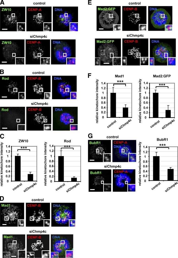
Figure 4.
Chmp4c is required for optimal localization of spindle checkpoint proteins to prometaphase kinetochores. Localization of ZW10 and Rod (A–C), Mad1 and Mad2:GFP (D–F), and BubR1 (G) in cells transfected with negative siRNA (control) or Chmp4c siRNA (siChmp4c). Relative green/red fluorescence intensity is shown, and values in the control were set to one. n > 200 kinetochores, 20 cells from three independent experiments. Error bars show the SD. ***, P < 0.001 compared with control. Student’s t test was used. In some Chmp4c-depleted cells, a few (typically two to six) prometaphase kinetochores exhibited detectable Mad1 or Mad2:GFP staining and are shown in insets with dotted lines (D and E). Insets show 1.7× magnification of kinetochores. Bars, 5 µm.

