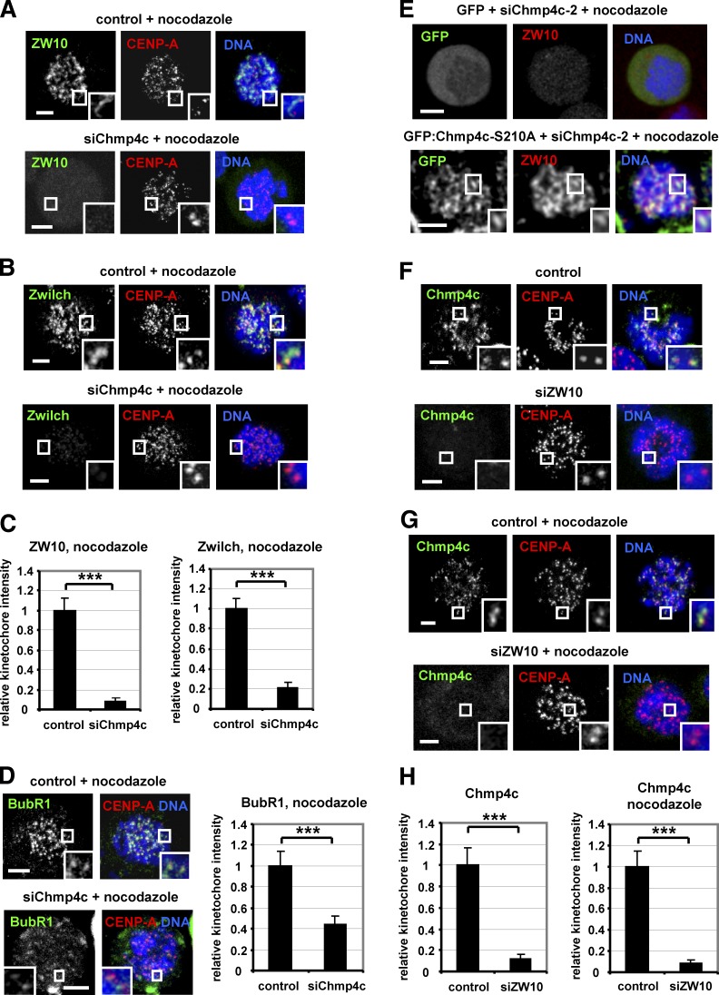
Figure 6.
Chmp4c and ZW10 localize to kinetochores in an interdependent manner. (A–D) Localization of ZW10, Zwilch, and BubR1. Cells were transfected with negative siRNA (control) or Chmp4c siRNA (siChmp4c) and treated with nocodazole for 4 h. (E) Cells expressing GFP or GFP: Chmp4c-S210A resistant to degradation by Chmp4c-2 siRNA (siChmp4c-2) were transfected with siChmp4c-2 and treated with nocodazole for 4 h. 20 cells from two independent experiments were analyzed. (F–H) Chmp4c localization. Cells were transfected with negative siRNA (control) or ZW10 siRNA (siZW10) in the absence or presence of nocodazole for 4 h. Relative green/red fluorescence intensity is shown, and values in the control were set to one. n > 200 kinetochores, 20 cells (C, D, and H) from three independent experiments. Insets show 1.7× magnification of kinetochores. Error bars show the SD. ***, P < 0.001 compared with the control. Student’s t test was used. Bars, 5 µm.

