Figure 2.
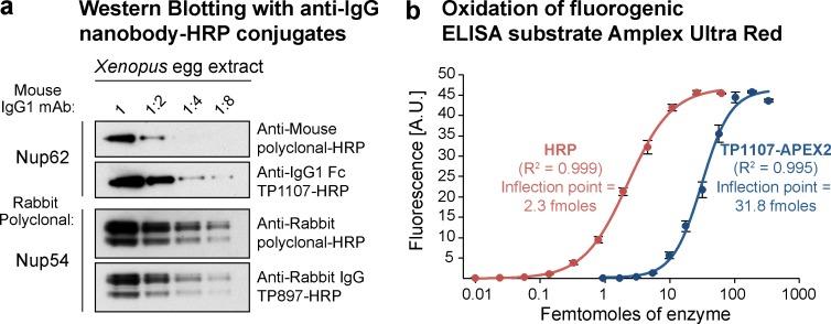
Application of peroxidase-linked anti-IgG nanobodies. (a) A twofold dilution series of Xenopus egg extract was blotted and probed with anti-Nup62 mouse IgG1 mAb A225. It was then decorated with HRP-conjugated goat anti–mouse polyclonal IgG (5 nM) or anti–mouse IgG1 Fc nanobody TP1107 (5 nM) and detected via ECL. Similarly, a rabbit polyclonal antibody targeting Nup54 was decorated with HRP-conjugated goat anti–rabbit polyclonal IgG or anti–rabbit IgG nanobody TP897 (5 nM). (b) Oxidation of the fluorogenic ELISA substrate Amplex Ultra Red. A dilution series of pure HRP or recombinant anti–mouse IgG1 Fc nanobody TP1107–APEX2 fusion was incubated with Amplex Ultra Red and H2O2. Oxidation leads to formation of the fluorescent compound resorufin. The data obtained were fit with a four-parameter logistic regression. The inflection points of the curves can be used to compare attainable sensitivity. A.U., arbitrary units. Error bars, mean ± SD (n = 3).

