
Figure 1.
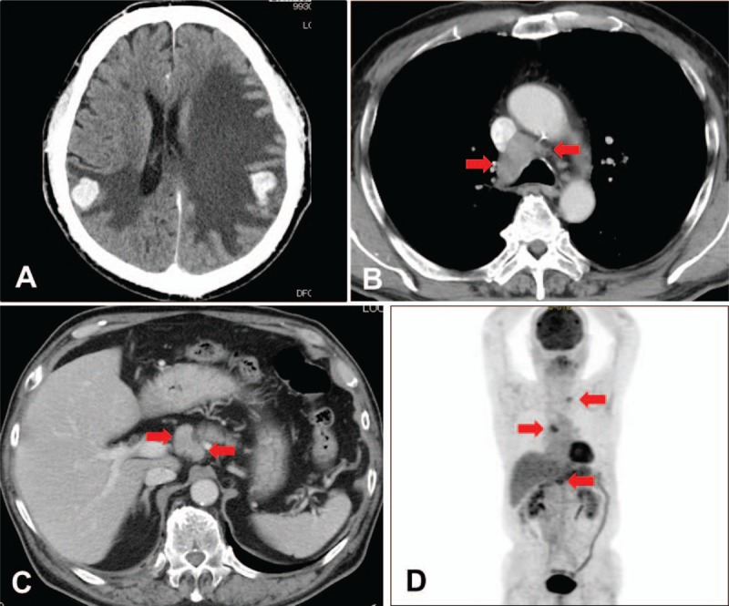
Initial computed tomography (CT) and [18F]-fluorodeoxyglucose (FDG) positron emission tomography (PET). (A) CT scan showed multiple nodules and edema in the bilateral cerebral hemispheres. (B) CT scan showed swelling of the mediastinal lymph nodes (arrowhead). (C) CT scan showed upper abdominal lymph node swelling (arrowhead). (D) FDG-PET scan demonstrated high FDG uptake at the same lymph nodes detected via CT scan (arrowhead).