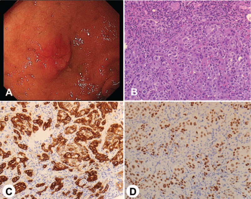
Figure 2.

(A) Upper gastrointestinal endoscopy showed a metastatic duodenal tumor. (B) A biopsy specimen showed histopathological findings of adenocarcinoma (hematoxylin and eosin stain, magnification ×200). (C) Immunohistochemical staining of the specimen showed CK7 positivity of the tumor cells (magnification ×200). (D) Immunohistochemical staining of the specimen showed TTF-1 positivity of the tumor cells (magnification ×200).
