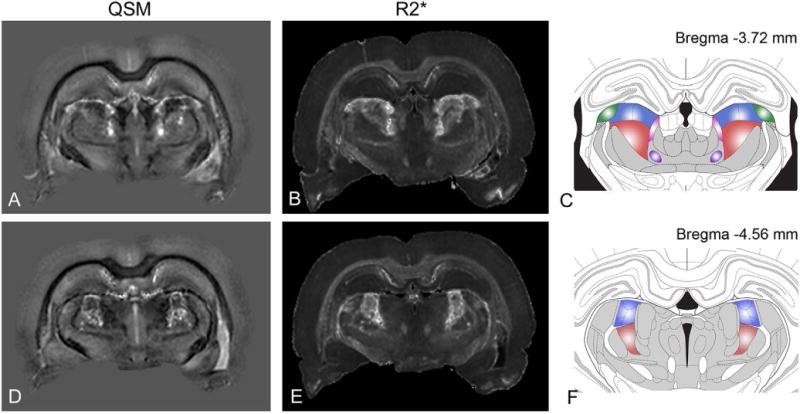
Figure 6.

Nuclei-specificity of thalamic lesions in the rat brain with pilocarpine-induced SE. QSM (A, D) and R2* (B, E) maps at two different coronal levels are compared with corresponding sections from the rat brain atlas (C, F) by Paxinos and Watson (2004). Major thalamic nuclei corresponding to the lesioned areas include: the lateral posterior nucleus (blue), dorsal lateral geniculate nucleus (green), posterior thalamic nuclear group (red), and the oval paracentral nucleus (purple).
