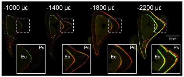
Figure 2.
Representative transverse sections of fluorochrome-labeled, loaded tibias illustrating increasing bone formation on periosteal (Ps) and endocortical (Ec) surfaces with increasing peak strain magnitude. Tibias in these groups were loaded 1200 cycles/day, 5 days/wk for 2 weeks. Mice were loaded days 1–5 and 8–12, injected with calcein on day 5 and alizarin on day 12, and euthanized on day 15. The sample in the −2200 με group has a small amount of periosteal woven bone.

