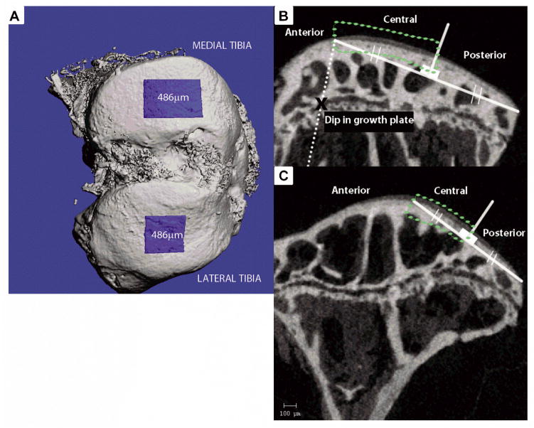
Figure 1.
A) Bird’s eye view of articular cartilage and subchondral bone region of interest B) Central region of articular cartilage was isolated within the medial tibia for thickness analysis. An x indicates the landmark (dip) in the tibial growth plate, through which a line (dotted) parallel to the tibial metaphyseal cortical bone was drawn. The intersection of this line with the tibial plateau surface was used to draw a secant line to the posterior edge of the plateau, which was bisected to isolate the central region C) Central region of articular cartilage within the lateral tibia isolated for thickness analysis. A secant line was drawn over the relatively flat portion of the posterior end of the plateau, and bisected to isolate the central region.

