
Figure 2. MeXis regulates Abca1 expression and function.
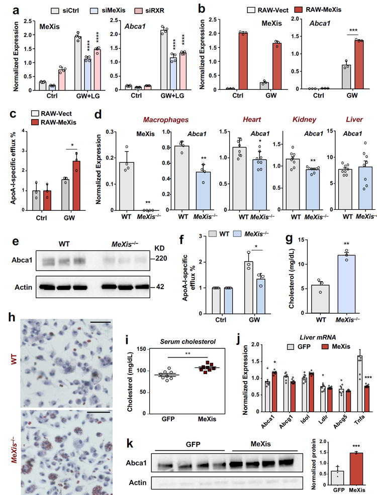
A. Real-time PCR analysis of MeXis and Abca1 expression from primary macrophages treated with the indicated siRNAs (50 nM) followed by either vehicle (Ctrl) or a combination of GW3965 (GW, 0.5 μM) and the RXR ligand LG268 (LG, 50 nM) for 36 h. Results are representative of four independent experiments. Values are means ± SD.**** P<0.0001 by Two-way ANOVA followed by multiple comparisons test (Sidak’s). B. Real-time PCR analysis of MeXis and Abca1 expression 10 days after stable overexpression of control vector (Vect) or MeXis in RAW cells treated with vehicle (Ctrl) or GW3965 (GW, 0.5 μM). Results are representative of three independent experiments. Values are means ± SD.*** P < 0.001 by two-sided student’s t-test. C. Cholesterol efflux in the presence of ApoA-I from RAW macrophages loaded with [3H]cholesterol (1.0 μCi/ml) and treated with the acyl-CoA:cholesterol O-acyltransferase inhibitor (2 μg/ml) and either with DMSO or LXR ligand (1 μM GW3965). ApoA-I-specific efflux represents percent radiolabelled cholesterol efflux in the presence of ApoA-I normalized to DMSO. Experiments were conducted in triplicate. Data are expressed as mean ± SD.* P<0.05 by two-sided student’s t-test.D. Real-time PCR analysis of MeXis and Abca1 expression in primary mouse macrophages (results are representative of four independent experiments; values are means ± SD) and of Abca1 expression in heart, kidney and liver of mice fed a western diet for 3 weeks (N = 8/group; values are means ± SEM).* P<0.05; ** P < 0.01 by two-sided student’s t-test.E. Western blot analysis of Abca1 levels in primary mouse macrophages of WT and MeXis−/− mice treated with GW (0.5 μM for 16 hours). Actin was used as a loading control. The experiment repeated twice with similar results. F. Cholesterol efflux in the presence of ApoA-I or HDL from WT or MeXis−/− macrophages loaded with [3H]cholesterol (1.0 μCi/ml) and treated with the acyl-CoA:cholesterol O-acyltransferase inhibitor (2 μg/ml) and either with DMSO or LXR ligand (1 μM GW3965). Experiments were conducted in triplicate. Data are expressed as mean ± SD. * P<0.05 by two-sided student’s t-test.G. Cholesterol content measured in peritoneal macrophages isolated from mice on western diet for 12 weeks (N = 3/group). ** P < 0.01 by two-sided student’s t-test. H. Oil-red-O staining of peritoneal macrophages isolated from WT or MeXis−/− mice and treated with oxidized LDL (100 μg/ml) for 72 h.The experiment was repeated 3 times with similar results.Scale bars, 50 μm. I. Total serum cholesterol levels in 10-week-old chow-fed male C57BL/6 mice transduced with adenoviral vectors encoding GFP control (Ad-GFP) or MeXis (Ad-MeXis) for 6 days (n = 8 per group). ** P < 0.01 by two-sided student’s t-test. J. GFP and MeXis expression in liver 6 days after transduction of mice with Ad-GFP or Ad-MeXis, respectively (n = 8 per group except Abcg1 n=7 per group).* P<0.05; *** P < 0.001 by two-sided student’s t-test. Data are expressed as mean ± SEM. K. Left, western blot analysis of Abca1 levels in liver from the mice in I (n = 4 per group). Right, quantification of protein levels normalized to actin. Data expressed as mean ± SD. *** P < 0.001 by two-sided student’s t-test.