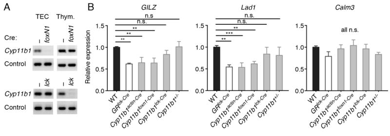
FIGURE 2. Reduced glucocorticoid-dependent gene expression in Cyp11b1foxN1-Cre thymocytes.

(A) Cre-mediated disruption of Cyp11b1. Genomic DNA from sorted TECs and DP thymocytes from WT and Cyp11b1foxN1-Cre (N1-Cre) and Cyp11b1lck-Cre (lck-Cre) mice was analyzed by PCR for the presence of CYP11B1 exon 4. Control primers were specific for the H-2A locus (41). One representative pair of three sets of mice for each Cre is shown. (B) mRNA levels of GC-sensitive and -insensitive genes in Cyp11b1foxN1-Cre thymocytes. Relative mRNA levels in thymocytes either freshly isolated or after 3 hr of treatment with 100 nM corticosterone were determined by RT-PCR. Significance was determined by 1-way ANOVA, followed by Fisher’s LSD multiple comparison (each mutant vs control, n =4 to 8). *P < 0.05, **P < 0.005, *** P < 0.0005.
