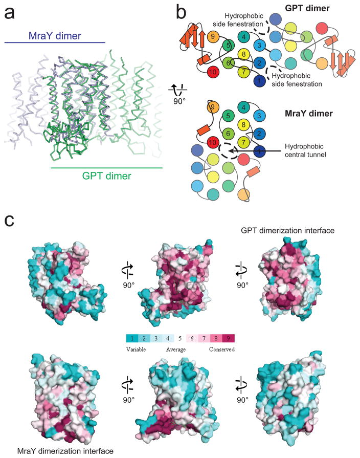
Figure 3. Divergence between GPT and MraY is underpinned by their different dimerization interfaces.
a, Superposition of hGPT (green) with MraYCB (blue) reveals strikingly different dimerization interfaces, despite the conserved fold of each protomer. b, The different dimerization interfaces of GPT and MraY result in altered membrane accessibility. Hydrophobic side fenestrations penetrate the GPT dimer, while a central hydrophobic tunnel occupies the MraY dimer. c, The interface for GPT dimerization is conserved only among GPT orthologue sequences but not among MraY sequences, and vice versa is true for MraY. Surfaces are colored in increasing conservation from cyan (least) to magenta (most). Sequence conservation was mapped onto the hGPT and MraYCB structures using 30 orthologue sequences for each alignment.

