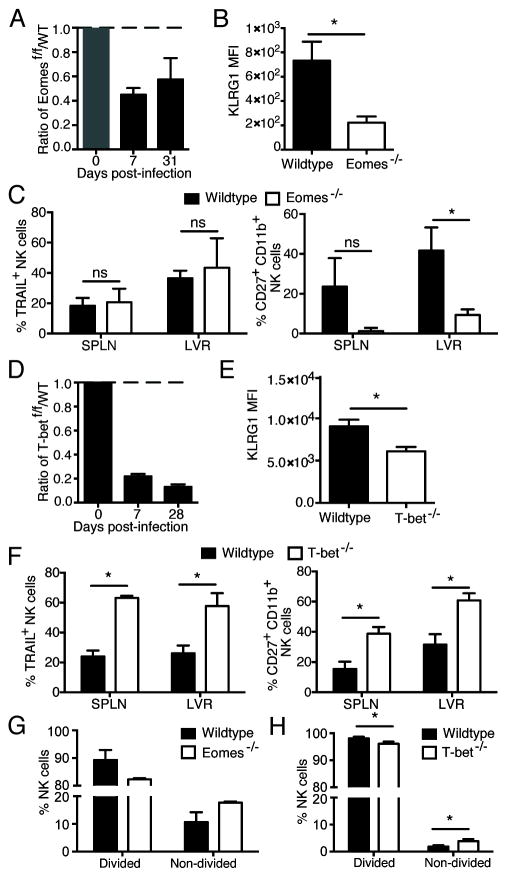
Figure 2. T-box transcription factors are necessary for the expansion of antigen-specific NK cells.
WT (CD45.1) and Eomesf/f x UbCre-ERT2 (CD45.2) or Tbx21f/f x UbCre-ERT2 (CD45.2) NK cells were co-transferred into Ly49h−/− mice and treated with tamoxifen or oil at day −4 PI. Mice were infected with MCMV at day 0 PI. (A and D) The populations of tamoxifen-treated Eomesf/f x UbCre-ERT2 or Tbx21f/f x UbCre-ERT2 NK cells and their WT counterparts relative to oil-treated populations are shown for each time point. (B and E) MFI of KLRG1 staining at day 7 PI in the spleen is shown. (C and F) Assessment of phenotypic markers at day 7 PI for the spleen and liver are shown. (G and H) Ly49H+ NK cells from WT and Eomesf/f x UbCre-ERT2 or Tbx21f/f x UbCre-ERT2 mice were labeled with CTV, transferred into a Ly49h−/− host which was treated with tamoxifen (or oil) at day −4 PI and infected with MCMV at day 0. Bar graph shows percentages of divided and non-divided NK cells at day 4 PI for each group. Data are mean ± SEM representative of two to three independent experiments with at least n=4 biological replicates per condition. * p < 0.05 and ns, not significant, paired Student t-test.

