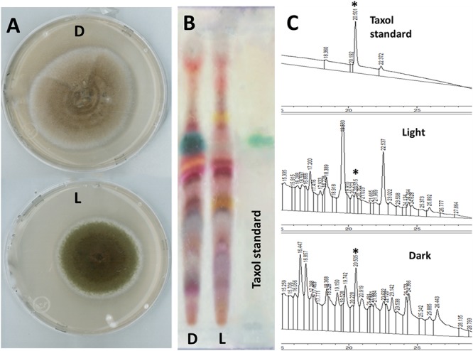
FIGURE 1.

Light pre-incubation inhibited fungal Taxol production. (A) Growth of Taxol-producing endophyte SSM001 fungus in light (L) and darkness (D) on PDA at 25°C for 1 week. (B) Detection of extracted fungal Taxol after inoculation for 3 weeks in liquid YPD broth on TLC-silica plates (10 μL sample, developing system chloroform: methanol; 5:0.5 and visualized using 0.5% vanillin/sulfuric acid reagent). (C) Detection and quantification of fungal Taxol by HPLC-UV when fortified with 5 ng standard Taxol. The peak area of each sample (10 μL injection volume) was measured at 233 nm. The quantification data display the mean of three replicates. The asterisk is the diagnostic peak of Taxol.
