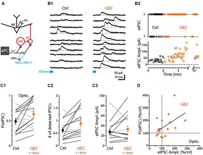
Figure 7.
Evidence for dSAC-mediated disinhibition of Mitral cells (MCs). (A) Schematic illustrating protocol for measuring disynaptic (CFF axon-to-GC-to MC) IPSCs in MCs. CFF stimulation was always conducted optogenetically in slices obtained from ntsr1-ChR2-YFP mice. IPSCs were recorded at depolarized holding potentials (0 mV or −45 mV). (B1) Consecutive current traces recorded in a MC at 0 mV. Under control conditions (left), disynaptic IPSCs were only occasionally observed, but a low concentration of gabazine (0.5 μM GBZ) enhanced the probability of the IPSCs. Light-stimulation was conducted by focusing the 40× objective in the GCL, 280 μm from the MCL. (B2) Time plots for the experiment in (B1), showing increases in the success-rate of light-evoked IPSCs due to low GBZ (top) and increased amplitude of the IPSCs (bottom). Each data point reflects one trial. (C) Summary of low GBZ effects on the probability of evoking an IPSC (C1), the number of evoked IPSCs (C2), and the amplitude of the IPSCs (C3). Plots reflect recordings from 16 MCs (averages of 24 trials per cell). MCs in which low GBZ increased the IPSC probability are shown with black lines in all plots; other MCs shown with light gray lines. *in (C1) p = 0.040, paired t-test, n = 16; *in (C2) p < 0.05, paired t-test, n = 16. (D) Low GBZ-effects on the probability of evoking an IPSC plotted as a function of the effect on IPSC amplitude. Line: linear regression fit, R2 = 0.23, p = 0.06.

