Figure 1.

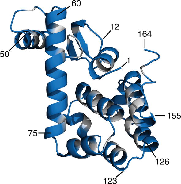
Crystal structure of T4 lysozyme (164 residues, PDB 2LZM, 1.7 Å resolution). Residues with low predicted labeling probabilities that were excluded from the test set are shown in grey. Several residues are labeled as guides to the eye.

Crystal structure of T4 lysozyme (164 residues, PDB 2LZM, 1.7 Å resolution). Residues with low predicted labeling probabilities that were excluded from the test set are shown in grey. Several residues are labeled as guides to the eye.