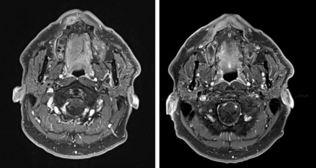
Figure 1.

Gadolium-enhanced MRI in a patient with a right labial localization of ACC visualized before (left panel) and 3 months after CIRT (right panel). The two measurements in the first MRI correspond respectively to the longitudinal (line 1:11 mm) and transversal diameters (line 2:15 mm) of the lesion. The tumor is no longer visible after therapy.
