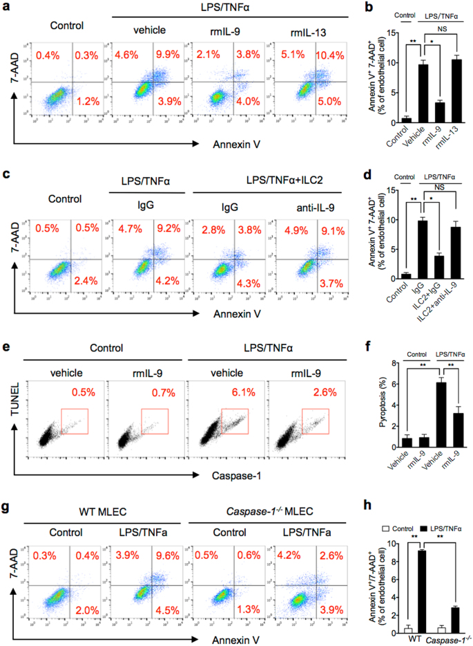
Fig. 5. ILC2-derived IL-9 protects MLEC from pyroptosis.
a, b Representative flow cytometry plots and quantitation of Annexin V/7-AAD staining of MLEC cultured with LPS/TNFα and with or without rmIL-9 or rmIL-13 for 24 h. c, d Representative flow cytometry plots and quantitation of Annexin V/7-AAD staining of MLEC co-cultured with ILC2 (1 × 10⁴ cells/well) and LPS (1 μg/ml) + TNFα (20 ng/ml) and with IgG or anti-IL-9 for 24 h. e, f Representative flow cytometry plots and quantitation of MLEC pyroptosis (caspase-1/TUNEL double-positive cells) with rmIL-9 (50 ng/ml), LPS, and TNFα for 24 h. g, h Representative flow cytometry plots and quantitation of Annexin V/7-AAD staining of MLEC from WT or Caspase-1−/− mice with LPS/TNFα for 24 h. MLECs were identified as CD31+ cells. Data shown are the mean ± SEM, n = 3–6 mice/group. *P < 0.05, **P < 0.01

