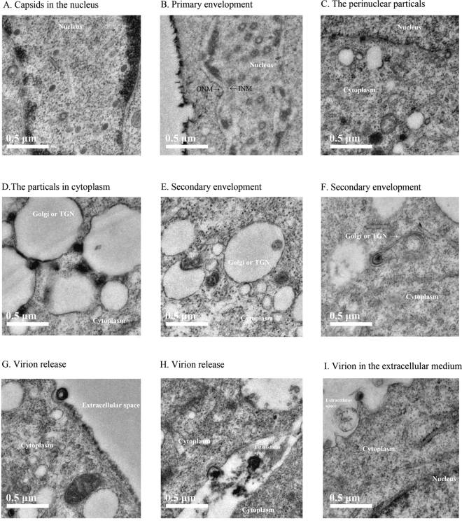
Figure 7.
Electron micrographs of the steps of gJ-deleted mutant virus lifecycle. (A) Capsids in the nucleus. (B) Primary envelopment, showing the close apposition of the particles and the inner nuclear membrane (INM). (C) Primarily enveloped particles present within the perinuclear space. (D) Particles in the cytoplasm, showing the close apposition of the particles and the Golgi or trans-Golgi network (TGN). (E) Initial steps of secondary envelopment. The unenveloped capsids in the cytoplasm interacted with TGN membrane and was being wrapped in these membranes. (F) Final steps of secondary envelopment. Enveloped particles were present with the TGN-derived membranes. (G) and (H) Virions release. Enveloped virions are transported to cell surface and released. (I) Virion in the extracellular medium.

