. 2018 Feb 12;27(1):45–56. doi: 10.5607/en.2018.27.1.45

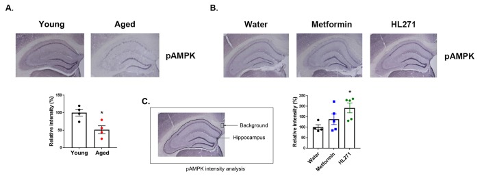
Fig. 4. AMPK activation in the mouse brain. (A) AMPK activation levels in young and aged mice. Upper: representative images of phospho-AMPK (pAMPK) expression in young and aged mice. Lower: quantification of pAMPK expression levels. Young (100±9.73, n=4) and aged (51.56±11.27, n=4). *p=0.0174, student's t-test, two-tailed. (B) Effects of HL271 on AMPK activation. Upper: representative images of pAMPK expression in the three groups. Lower: quantification of pAMPK expression levels. One-way ANOVA; F (2, 11)=4.142, *p=0.0456. Post hoc Bonferroni's multiple comparisons unveiled significant differences between the water (100.4±11.72, n=4) and HL271-treated mice (192.4±23.52, n=5), *p=0.0484. There was no significant difference between the water and metformin-treated group (138.6±25.45, n=5), p=0.7907. (C) Image describing the pAMPK intensity analysis. Using the freehand selection tool in ImageJ, a line was drawn to select the whole hippocampus, and the mean intensity was measured. A square box of a fixed size was used to measure the background intensity for each slice. Data are presented as the mean±SEM.

Fig. 4