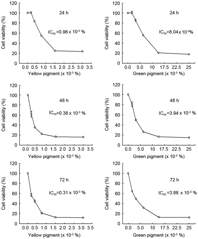
Figure 3.
Cell viability analysis in green or yellow pigment-treated DLD-1 cells. DLD-1 cells were plated in 60 mm culture dishes at 80% confluence and treated with the indicated concentrations of pigment for 24 h. Following treatment, the MTT reagent (0.5 mg/ml) was added to the cells for 2 h at 37°C, and the cells were lysed with dimethyl sulfoxide. Absorbance was evaluated at 595 nm. IC50, half-maximal inhibitory concentration.

