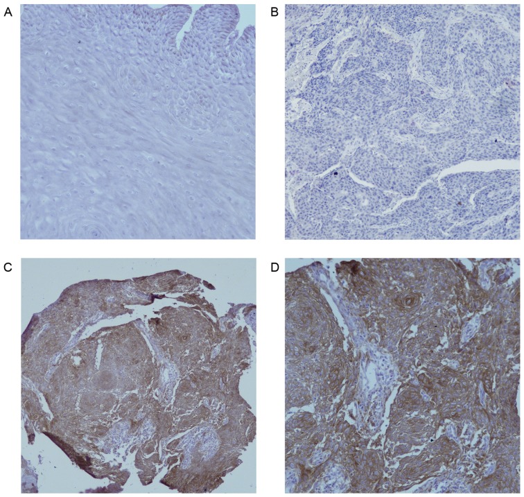
Figure 1.
PD-L1 expression in EC and normal esophageal epithelium. (A) No PD-L1 staining in normal esophageal epithelium (magnification, ×200). (B) Negative expression of PD-L1 in EC (magnification, ×200). (C) Positive expression of PD-L1 in the cytoplasm and membrane of EC cells (magnification, ×100). (D) Positive expression of PD-L1 in the cytoplasm and membrane of EC cells (magnification, ×200). PD-L1, programmed death ligand 1; EC, esophageal carcinoma.

