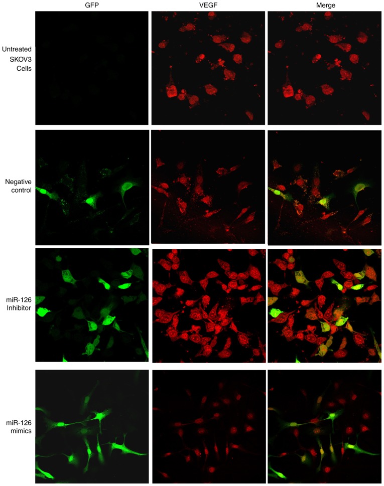
Figure 3.
Immunofluorescence staining demonstrated suppression of VEGF by miR-126 in transfected cells. Cells were grown on coverslips overnight and subsequently subjected to immunostaining with anti-VEGF antibody. VEGF-positive cells were stained red, whereas GFP-positive cells were stained green. Merged images present VEGF and GFP. miR-126, microRNA-126; VEGF, vascular endothelial growth factor; GFP, green fluorescent protein.

