
Fig. 1.
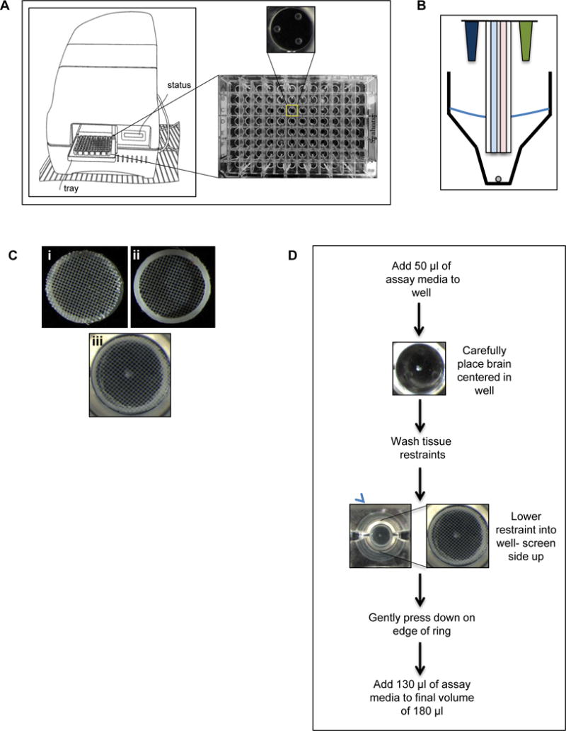
Design and implementation of a tissue restraint for analyzing metabolism using the XFe96 metabolic analyzer.
A) Cartoon of the XFe96 metabolic analyzer and cell plate. Zoomed in view of the bottom of a single well depicting the three raised spheres that the sensor probes rest on. B) Cartoon of the XFe96 probes (blue: oxygen consumption detection, pink: extracellular acidification detection) with injection ports (dark blue and green), and tissue being measured (grey circle). C) Images of tissue restraint, i) nylon screen facing up (correct orientation), ii) bottom view of restraint with plastic ring facing up, and iii) tissue restraint in well of XFe96 cell plate in correct orientation holding down a Drosophila larval brain. D) Protocol for analyzing Drosophila brain with the XFe96 metabolic analyzer using the tissue restraint.