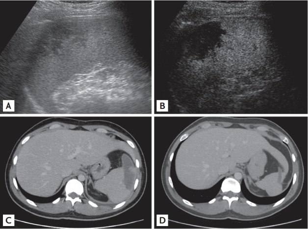
Figure 1.

Abdominal imaging in the patient. (A) Ultrasound sonography showed a peripheral wedge-shaped hypoechoic area in an enlarged spleen. (B) Late-vascular phase (i.e., 60 seconds after injection) of contrast-enhanced ultrasound sonography with Sonazoid (Daiichi Sankyo, 0.015 mL/kg) showed a non-enhanced infarcted area as a wedge-shaped area. (C) A portal venous phase computed tomography (CT) taken on the admission day showed splenomegaly and a wedge-shaped hypoenhancement area. (D) A portal venous phase CT after 2 months of discharge revealed decreased splenic infarcted areas accompanied by localized atrophic changes in the infarcted areas.
