Figure 2.
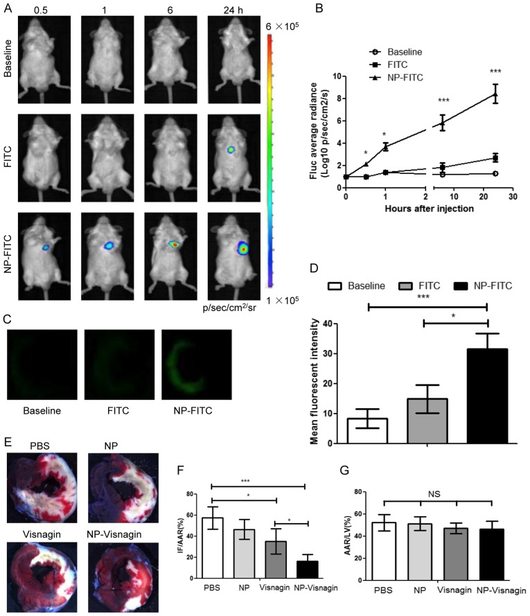
NP-visnagin targeted to the reperfused myocardium and exerted cardioprotective effects. (A) Following the intravenous injection of PBS (baseline), FITC or NP-FITC, in vivo BLI was performed at each time point. (B) Quantitative analysis of BLI data for NP-FITC compared with FITC alone. (C) Fluorescence images of heart cross-sections at 12 h after the intravenous injection of PBS, FITC or NP-FITC. (D) Quantification of the fluorescence intensity of the non-ischemic myocardium and AAR. (E) Representative stereomicrographs of heart sections double stained with TTC and Evans blue at 72 h after intravenous injection. (F) Quantitative analysis of IF/AAR. (G) Quantitative analysis of AAR/LV. *P<0.05; ***P<0.005. NP, nanoparticles; FITC, fluorescein isothiocyanate; BLI, bioluminescence imaging; TTC, 2,3,5-triphenyltetrazolium chloride; IF, infarct area; AAR, area at risk; LV, left ventricle; NS, not significant.

