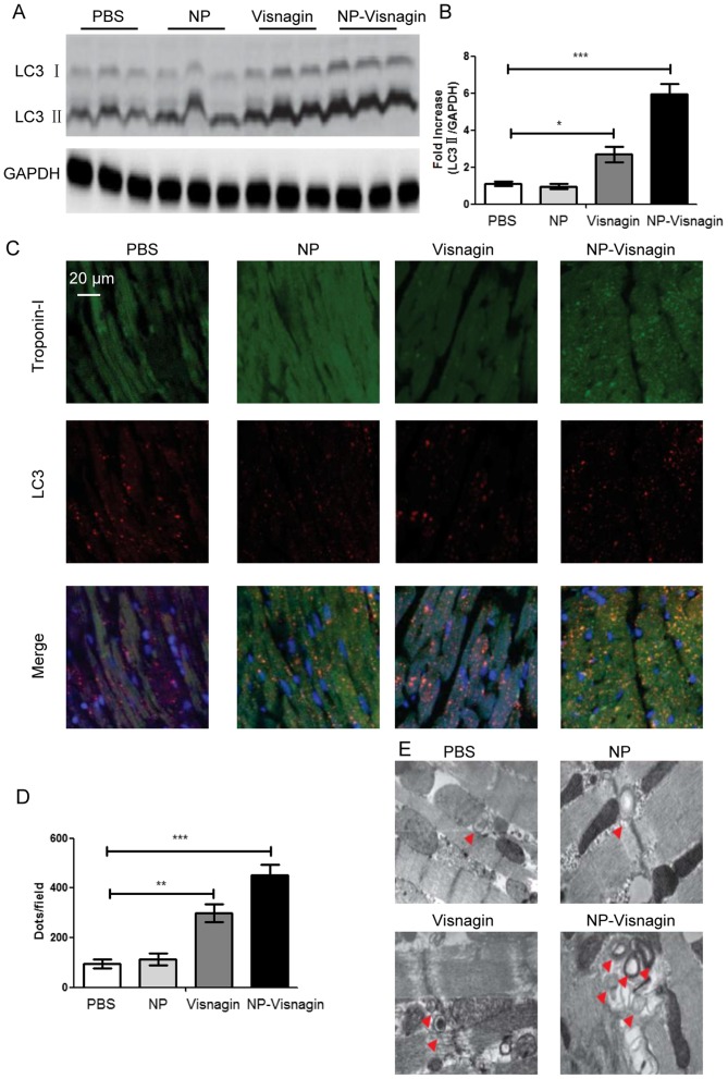
Figure 4.
NP-visnagin enhanced the rate of autophagy in the ischemic region after ischemia/reperfusion. (A) Western blotting was performed for the detection of autophagic flux; LC3II was detected in protein harvested from the treated hearts at 24 h after intravenous injection. GAPDH was detected as an internal standard. (B) Quantification of the expression of LC3II relative to GAPDH. (C) Representative images of the immunofluorescence detection of Troponin-I and LC3. Green dyeing represents Troponin-I, indicative of cardiomyocytes, whereas red dots represent autophagosomes. (D) Quantification of LC3 immunofluorescence. (E) Detection of double membrane autophagosomes with transmission electron microscopy. *P<0.05; **P<0.01; ***P<0.005. NP, nanoparticles; LC, light chain.

