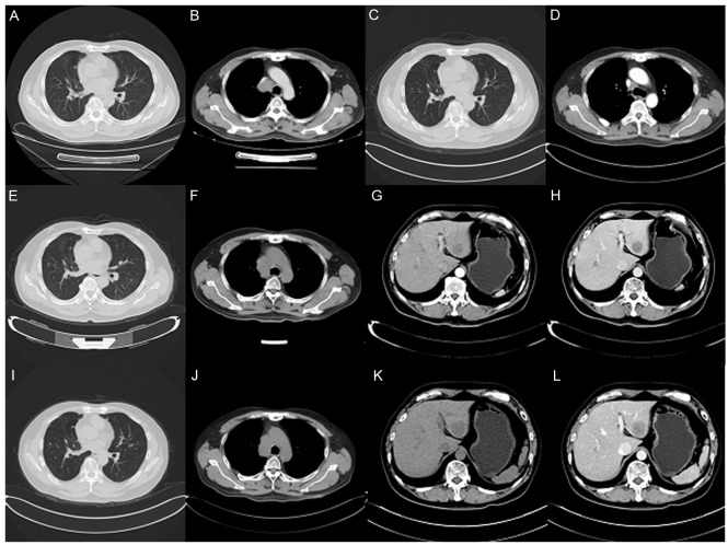
Figure 1.
Computed tomography scans of the patient. (A and B) Aug 21, 2015: Multiple lung metastases appeared and the right lung hilar lymph node appeared enlarged, as visualized in the (A) pulmonary and (H) mediastinal windows. (C and D) Dec 21, 2015: Regression of lung metastatic neoplasm and notable shrinking of the right mediastinal lymph node, as visualized in the (C) pulmonary and (D) mediastinal windows. (E and F) Mar 16, 2016: Lung metastasis and hilar lymph node was markedly enlarged, as visualized in the (E) pulmonary and (F) mediastinal windows. (G and H) Mar 16, 2016: Emerging new liver metastasis as visualized in the (G) arterial and (H) portal phases. (I and J) May 10, 2016: Lung metastasis and enlarged mediastinal lymph node, as visualized in the (I) pulmonary and (J) mediastinal windows. (K and L) May 10, 2016: Liver metastasis as visualized in the (K) arterial and (L) portal phases.

