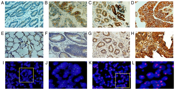
Figure 2.
Variable expression of RAB1A and HER-2 in human GAC tissue samples. The variable expression of RAB1A and HER-2 in human GAC tissue samples was assayed by (A-H) IHC and (I-L) FISH. (A) Negative membrane staining for HER-2 in GAC epithelium (0+). (B) Weak membrane staining in >10% tumour cells (1+). (C) Moderate complete membrane staining in >10% tumour cells (2+). (D) Strong complete membrane staining in >10% tumour cells (3+). (E) Cytoplasmic staining for RAB1A in normal gastric epithelium (negative). (F) Cytoplasmic staining for RAB1A in tumour cells (weakly positive). (G) Cytoplasmic staining for RAB1A in tumour cells (moderately positive). (H) Cytoplasmic staining for RAB1A in tumour cells (strongly positive). (I and J) FISH assay representing negative HER-2 amplification. (K and L) FISH assay representing positive HER-2 amplification (magnification, ×100). HER-2, human epidermal growth factor receptor 2; CAG, gastric adenocarcinoma; IHC, immunohistochemistry; FISH, fluorescence in situ hybridization.

