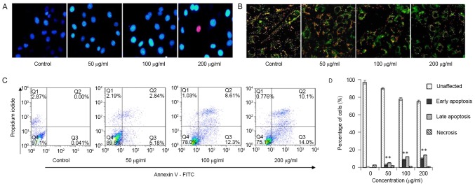
Figure 4.
Steviol induced apoptosis of U2OS cells. (A) Photomicrographs of Hoechst 33342 staining. (B) Concentration-dependent effect of steviol on mitochondrial membrane potential of U2OS cells. As steviol concentration increased, the green fluorescence intensity increased. (C) U2OS cells were treated with steviol for 48 h and stained with Annexin V-propidium iodide. (D) The percentage of apoptotic cells was measured by flow cytometry analysis *P<0.05 and **P<0.01 vs. untreated cells. FITC, fluorescein isothiocyanate.

