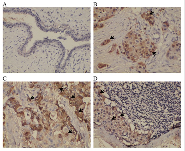
Figure 2.

Expression of protein S100-A8 in non-metastatic breast tumor, primary breast tumor, paired metastatic lymph node and control benign breast disease tissues, as detected by immunohistochemical staining. (A) BBD tissue exhibited light or negative expression of protein S100-A8 in epithelial cells. Strong to moderate protein S100-A8 cytoplasm staining was observed in (B) NMBT and (C) PBT. Light protein S100-A8 cytoplasm staining was observed in (D) PMLN. Sporadic and light-moderate expression of protein S100-A8 was identified in the nuclei of cancerous cells (identified by a black arrow). Staining, 3,3′-Diaminobenzidine; magnification, ×200. CT, cancerous tissue; LT, lymph tissue.
