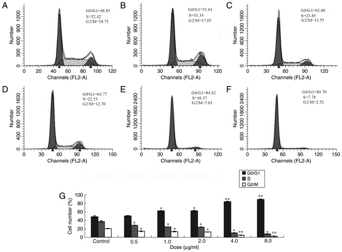
Figure 3.
Cell cycle of MCF-7 cells following treatment with different doses of ailanthone. MCF-7 cells were treated with different doses of ailanthone for 48 h and the cell cycle distribution was measured by flow cytometry. (A) Control (0 µg/ml) and (B) 0.5 µg/ml, (C) 1.0 µg/ml, (D) 2.0 µg/ml, (E) 4.0 µg/ml and (F) 8.0 µg/ml ailanthone. (G) Cell cycle distribution of MCF-7 cells. Data are presented as the mean ± standard deviation (n=3). *P<0.05 and **P<0.01 vs. the respective control group.

