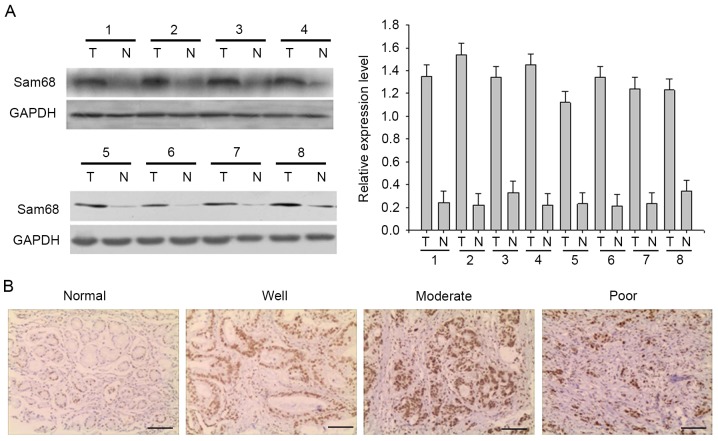
Figure 1.
Expression of Sam68 in human gastric cancer. (A) Western blot analysis revealed high Sam68 protein levels in gastric cancer. GAPDH was assessed as a loading control. The bar chart demonstrates the ratio of Sam68 protein to GADPH using densitometry analysis. The data are presented as the mean ± standard deviation of three independent experiments. (B) Paraffin-embedded gastric (normal) or gastric cancer tissue sections (well, moderate, poor) were stained with antibodies against Sam68, and then counterstained with hematoxylin (Scale bar, 100 µm). Sam68, Src-associated in mitosis of 68 kDa.

