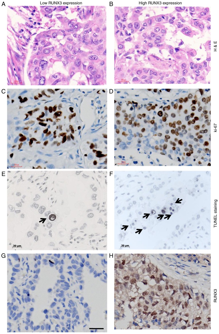
Figure 5.
Cellular dynamic parameters between patients with NSCLC and different expression levels of RUNX3. H&E staining for NSCLC tissue with (A) low and (B) high level of RUNX3. Immunostaining for Ki-67 in NSCLC tissue with (C) low and (D) high level of RUNX3. TUNEL staining for apoptotic cells in NSCLC tissue with (E) low and (F) high level of RUNX3, characterized by chromatin condensation and nuclear fragmentation, which were accompanied by cell swelling, reduction in cellular volume (pyknosis) and retraction of pseudopodes. Formation of crescentic caps of condensed chromatin at the nuclear periphery, and formation of apoptotic bodies could also be observed (indicated by arrows). (G) Negative staining for RUNX3; (H) positive staining for RUNX3 in NSCLC. Magnification, ×400; scale bars=20 µm. NSCLC, non-small cell lung cancer; RUNX3, runt-related transcription factor 3; H&E, hematoxylin and eosin; TUNEL, terminal deoxynucleotidyl transferase mediated dUTP-biotin nick end labelling.

