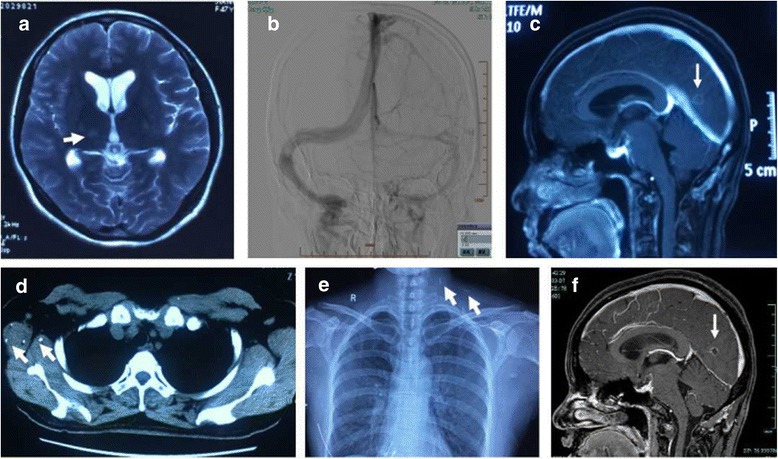
Fig. 1.

a The T2-weighted image showed a high signal of right thalamus. b DSA showed the probable underdevelopment on right lateral sinus while the venous system was smooth. c Cerebral MRI with contrast showed several small hyperintense lesions involving right cerebellar hemisphere and bilateral occipital, with ring enhancement of gadolinium. d The pulmonary CT showed multiple densities in the chest wall soft tissue. e Chest radiographs showed multiple calcification on the left side of the neck and right soft tissue of armpit chest wall. f The secondary MRI showed the nearly same small lesions with ring enhancement in the brain
